
Major advances have recently been made that have considerably advanced our understanding of the brain at the level of its cellular structure.
It has been known for over a century that the basic cell of the brain and the nervous system, the neuron, functions electrically. In the human brain there are some hundred billion neurons. To put this number in some perspective, imagine that an area covered by a similar number of house bricks would be over 500 square miles in size.
When a neuron receives electrical inputs from other neurons, electrical potential above a certain threshold “fires” off a signal that is transmitted to other neurons. The interaction between neurons takes place through a tiny space between them called a synapse, with chemicals known as neurotransmitters that can enhance or suppress the connections.
Although synapses can be studied separately under an electron microscope it is only recently that it has become possible to study the thousands of synaptic connections that each neuron can make with surrounding neurons in very minute detail. Up to 17 different proteins that are involved in synapses can be highlighted by binding differently coloured fluorescent molecules to them.
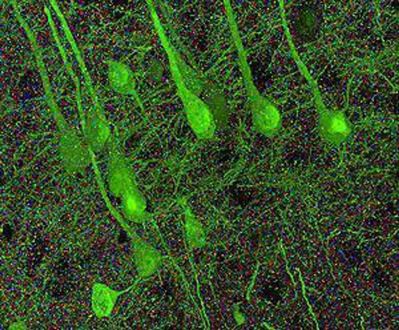 Synapses in the cortex of a mouse. The large green
blobs are neurons and the multicolored dots represent
separate synapses. ( Image courtesy of Stephen Smith
and Stanford University Medical Center)
A team at Stanford University School of Medicine, led by Professor Stephen Smith, used a technique called array tomography to create images like the one shown. Each tiny coloured dot in the picture shows a synapse, with different proteins shown by their colour. Synapses can now be identified by the combination of proteins they are composed of, enabling a classification of up to 12 types of synapses.
Smith pointed out in the press report of their findings [1] that in the human cerebral cortex alone—the rippling outer layer of the brain that is mainly responsible for giving humankind their special qualities—there are more than 125 trillion synapses. When one considers that a typical cortex if spread out is two to four millimetres thick over an area of 1.5 square metres, it can be understood just how densely the synaptic connections are packed together.
In the past attempting to map the labyrinthine circuitry of the cortex was impossible and picking out synapses was beyond the resolution of the most powerful microscopes. “Now we can actually count them and, in the bargain, catalog each of them according to its type,” Smith explained.
The Smithlab team at Stanford used a slab of tissue from a mouse cerebral cortex (which has similar neurons and synapses to those of the human cortex although far fewer in total). They cut this slab into slices that are only 70 nanometers thick (100 nanometers is about a thousandth of the thickness of a human hair). These slabs are suitably treated to attach flourescent molecules to the various proteins and high resolution images created both with light and a Scanning Electron Microscope. Computer tomography methods are then used to put the images of the slices together. The resulting video showing navigation through a tiny volume of a mouse cortex can be viewed on YouTube [2].
Attempting to explain the incredible complexity of the brain that is revealed, Smith said, “One synapse, by itself, is more like a microprocessor—with both memory-storage and information-processing elements—than a mere on/off switch. In fact, one synapse may contain on the order of 1,000 molecular-scale switches. A single human brain has more switches than all the computers and routers and Internet connections on Earth,” he said.
It is not only the complexity of synapses that is now beginning to be understood but also the changeability of these connections. Studying synapses from laboratory specimens does not bring this out, but recently developed techniques allow them to be studied in a living brain. For example the laboratory led by Professor Charles D. Gilbert at Rockefeller University have been able to use a viral labelling method to attach fluorescent proteins to individual neurons in the somatosensory cortex of a living mouse (the part of the cortex receiving information from the body's touch and positional sensors) [3].
Although it is well known that synapses develop rapidly in early life, the Gilbert lab were able to show that brain circuits can be modified by experience in adult animals. After removing a whisker from a mouse they could image the proteins of individual synapses in the living brain by a technique known as two photon microscopy. They showed that the neurons receiving inputs from the mouse’s other whiskers changed their relationship as new circuits rapidly developed.
Perhaps even more fundamental than developments in understanding neurons and their synaptic connections is the change in scientists’ attitude towards the cells that make up some 85 percent of the brain other than neurons—so-called glia cells. It was long thought that glia (which means “glue” ) cells played a supporting role in the brain, holding together the neurons. In the last few years that view has changed as glia have been found to carry out a range of functions essential to the life of the brain [4].
Glia functions can be divided into three categories. Astrocyte glia (so-called because they resembled stars to early anatomists) fill the spaces between neurons, providing energy sources and maintaining the chemical surroundings of neurons in which they can fire electrical impulses and communicate chemically through synapses.
Then there are microglia that perform the role of an immune system within the brain. Since the brain is sealed off by a barrier from the rest of the body it needs its own immune system, killing bacteria and healing the brain after injury.
Thirdly there are glia that form the electrical insulation, called myelin, of the nerve fibres (axons) that in turn form the roots of neurons. Axons tightly bundled together and insulated with myelin form the “white matter” that makes up half of the brain.
Research into the different types of glia is now a high priority because it seems that they play a much more dynamic role in the brain than was once thought. White matter, for example, has been shown to change during experience, with myelin being formed by electrical impulses in the axons. As the speed of electrical transmission can increase 50 times in an axon insulated by myelin compared to one that is bare, this has a major impact on neural processing.
There is much controversy surrounding the role of astrocyte glia [5]. It seems that these glia, which have thousands of bushy tendrils that are close to the synapses, both respond to and influence synaptic activity. But it is now being claimed that astrocytes can respond to activity at one synapse and communicate with other synapses—so-called “gliotransmission.” Since any such glia can make tens of thousands of connections with surrounding cells, this would vastly increase the scale of brain processing far beyond the already amazingly complex situation of neurons and synapses explained by Professor Smith.
Apart from the huge medical potential of such fundamental brain research, with possibilities of cures for a range of degenerative diseases if adequate resources were put at the disposal of researchers, its philosophical significance for Marxists should not be underestimated. Marxist materialism has always insisted that mind and consciousness can only be understood in the context of the historical development of human society. One cannot take a simplistic view of thought that merely equates it with the electrical and chemical processes in a single brain. Nevertheless humans do think “with the help of the brain” as Lenin put it, and the almost unimaginable complexity of this organ that is now being uncovered helps to sweep aside all the arguments that a spiritual source must be necessary to explain the wonders of the human mind.
[1] http://www.sciencedaily.com/releases/2010/11/101117121803.htm
[2] http://www.youtube.com/watch?v=pNaQ2HAj1rY&feature=related
[3] http://www.sciencedaily.com/releases/2010/06/100615191647.htm
[4] Special section on Glia in Science, Vol 330, 5 November, 2010.
[5] “Settling the great glia debate”, Nature, Vol 468, 11 November, 2010, pp 160-162.